For citation:Sergeev A.Yu., Sergeev Yu.V. Dermatophytosis // BC. 2003. No. 15. P. 845
MMA named after I.M. Sechenov, National Academy of Mycology, Moscow
Causative agents Dermatophytes are mold fungi - ascomycetes of the family Arthodermataceae (order Onygenales) belonging to three genera - Epidermophyton, Microsporum and Trichophyton... In total, 43 types of dermatophytes are known, of which 30 are pathogens of dermatophytosis.
The main causative agents of mycoses in Russia, according to our data, are, in order of occurrence, T. rubrum (fig. 1), T. mentagrophytes (fig. 2), M. canis (fig. 3).
Figure: 1. Micromorphology of T. rubrum

Figure: 2. Micromorphology of T. mentagrophytes var. interdigitale

Figure: 3. Micromorphology of M. canis
Dermatophytes are called geophilic, zoophilic or anthropophilic, depending on their usual habitat - soil, animal or human. Members of all three groups can cause human diseases, but their different natural reservoirs determine the epidemiological characteristics - the source of the pathogen, the prevalence and geography of the areas (Table 1).

Although many geophilic dermatophytes can cause infection in both animals and humans, the most common natural habitat for these fungi is soil. Members of the zoophilic and anthropophilic groups are believed to have evolved from these and other soil-inhabiting saprophytes capable of destroying keratin. Zoophilic organisms can sporadically be transmitted to humans if they have an affinity for human keratin. Transmission occurs through direct contact with an infected animal, or through objects that get the wool and skin scales of these animals. Infections often occur in rural areas, but at present, the role of domestic animals is especially important (especially with infection M. canis). Many members of the zoophilic group are named after their animal hosts. The general epidemiological characteristic of zoonotic and anthroponous dermatophytosis is high contagiousness. Dermatophytosis is perhaps the only contagious infection among all human mycoses.
Although many geophilic dermatophytes can cause infection in both animals and humans, the most common natural habitat for these fungi is soil. Members of the zoophilic and anthropophilic groups are believed to have evolved from these and other soil-inhabiting saprophytes capable of destroying keratin. Zoophilic organisms can sporadically be transmitted to humans if they have an affinity for human keratin. Transmission occurs through direct contact with an infected animal, or through objects that get the wool and skin scales of these animals. Infections often occur in rural areas, but at present, the role of domestic animals is especially great (especially with infection). Many members of the zoophilic group are named after their animal hosts. The general epidemiological characteristic of zoonotic and anthroponous dermatophytosis is high contagiousness. Dermatophytosis is perhaps the only contagious infection among all human mycoses.The nature of infections caused by anthropophilic dermatophytes is, as a rule, epidemic. The main increase in the incidence is provided by anthropophilic species. Currently, anthropophilic dermatophytes can be found in 20% of the total population, and the infections they cause are the most common mycoses. According to our epidemiological study, there is an increase in the incidence of dermatophytosis.
Pathogenic properties and pathogenesis All dermatophytes have keratinolytic activity, i.e. capable of degrading animal and / or human keratin. The activity of keratinases and proteolytic enzymes in general is considered the basis of the pathogenic properties of dermatophytes. Keratinases themselves are capable of degrading not only keratin, but also other animal proteins, including collagen and elastin. The activity of keratinases is not the same in different dermatophytes. The most active is T. mentagrophytes, very moderate - T. rubrum... The ability to degrade different types of keratin in general corresponds to the localization of dermatophyte infection. So, E. floccosum - a species with low keratinolytic activity - does not affect hair.
The introduction of the pathogen colony into the epidermis is provided by both keratinolytic activity and hyphal growth. Like molds, dermatophytes have a specialized apparatus for the directed growth of hyphae. It is directed to the points of least resistance, as a rule, to the joints between adjacent cells. The penetrating hyphae of dermatophytes are traditionally considered special perforating organs. It is still unclear whose role in the invasive process is more important - keratinases or directed growth pressure.
The depth of advancement of the fungal colony in the epidermis is limited. With skin infections, dermatophytes rarely penetrate deeper than the granular layer, where natural and specific protective factors meet them. Thus, dermatophyte infection covers only inanimate, keratinized tissues.
The available data on the protection factors of the macroorganism in dermatophytosis cast doubt on the point of view of some authors that with this infection lymphohematogenous spread of the pathogen or its occurrence in non-keratinizing tissues washed by blood occurs. Deep forms of dermatophytosis are described in patients with severe deficiency of one or more resistance factors.
Classification The international classification of mycoses, adopted in ICD-10, is based on the principle of localization (Table 2). This classification is convenient from a practical point of view, but does not take into account the etiological features of dermatophytosis in some localizations. At the same time, the variants of etiology determine the epidemiological characteristics and the need for appropriate measures, as well as the characteristics of laboratory diagnosis and treatment. In particular, representatives of the genera Microsporum and Trichophyton have unequal sensitivity to some antimycotics.

For a long time, the generally accepted classification in Russia was the one proposed by N.D. Sheklakov in 1976. In our opinion, a reasonable and acceptable compromise is the use of the ICD classification with clarification, if necessary, of the etiology of the pathogen or its equivalent. For example: dermatophytosis of smooth skin ( tinea corporis B35.4) caused by T. rubrum (syn. rubrophytosis of smooth skin). Or: dermatophytosis of the scalp (B35.0 favus / microsporia / trichophytosis).
The term "dermatomycosis", with which they sometimes try to replace the commonly used name of dermatophytosis, in our opinion, is inappropriate and cannot be the equivalent of dermatophytosis. Dermatomycoses are fungal infections of the skin in general, i.e. and candidiasis, and versicolor versicolor, and many mold mycoses.
Dermatophytosis of the scalp Abroad, the following clinical and etiological forms are distinguished tinea capitis: 1) ectotrix infection. Called Microsporum spp. (anthropozoonotic microsporia of the scalp); 2) endotrix infection. Called Trichophyton spp. (anthroponous trichophytosis of the scalp); 3) favus (scab). Called T. shoenleinii; 4) kerion (infiltrative-suppurative dermatophytosis).
The most common of the listed infections in Russia is microsporia ... The main causative agent of dermatophytosis of the scalp in Russia and Eastern Europe is Microsporum canis... The number of registered cases of microsporia in recent years has been up to 100 thousand per year. The incidence of pathogens of anthroponous microsporia ( M. ferrugineum) and trichophytosis ( T. violaceum), common in the Far East and Central Asia, should be recognized as sporadic.
The classic picture of microsporia is usually represented by one or more rounded foci with fairly clear boundaries, from 2 to 5 cm in diameter. The hairs from the foci are dull, brittle, light gray, dressed in a white sheath at the base. Hair loss above the surface of the skin explains why the lesions appear to be clipped, consistent with the name ringworm. The skin in the center is slightly hyperemic and edematous, covered with small grayish scales. The specified clinical picture corresponds to the name "versicolor of gray spots".
For trichophytosis of the scalp multiple isolated small (up to 2 cm) foci are characteristic. Breaking off of hair at the level of the skin is typical, leaving a stump in the form of a black dot protruding from the orifice of the follicle (“lichen of blackheads”).
Classic painting favusa characterized by the presence of scooters ( scutula, lat. shield) - a crust of dirty gray or yellow. The formed scutula is a dry saucer-like crust, from the center of which hair emerges. Each scutula consists of a mass of hyphae glued together by exudate, i.e. is essentially a fungal colony. In advanced cases, the scutulas merge, covering most of the head. A solid crust with a favus resembles a honeycomb, which is due to the Latin name of the disease. With a widespread favus, an unpleasant, "mouse" (barn, cat) smell comes from the crusts. Currently, favus is practically not found in Russia.
The infiltrative-suppurative form of microsporia and trichophytosis is characterized by pronounced inflammation with a predominance of pustules and the formation of large formations - kerions. Kerion - a painful dense focus of erythema and infiltration - has a convex shape, looks bright red or cyanotic, with clear boundaries and a bumpy surface, covered with numerous pustules and erosions, often hidden under purulent hemorrhagic crusts. Characterized by dilated follicular orifices, from which yellow pus is released when pressed. A similar picture is compared with honeycombs ( kerion). Kerion is often accompanied by general symptoms - fever, malaise, headache. Painful regional lymphadenitis (usually posterior cervical or posterior nodes) develops.
Dermatophytosis of nails Onychomycosis affects at least 5-10% of the Russian population, and over the past 10 years, the incidence has increased 2.5 times. Onychomycosis on the feet occurs three to seven times more often than on the hands. Dermatophytes are considered the main causative agents of onychomycosis in general. They account for up to 70-90% of all fungal nail infections. The causative agent of onychomycosis can be any of the dermatophytes, but most often there are two types: T. rubrum and T. mentagrophytes var. interdigitale. T. rubrum - the main causative agent of onychomycosis in general.
Allocate three main clinical forms of onychomycosis: distal-lateral, proximal and superficial, depending on the site of introduction of the pathogen. The most common form is distal. In this case, the elements of the fungus penetrate into the nail from the affected skin in the area of \u200b\u200bthe broken connection of the distal (free) end of the nail and skin. The infection spreads to the root of the nail, and its advance requires the superiority of the growth rate of the fungus over the rate of natural growth of the nail in the opposite direction. Growth of the nail slows down with age (up to 50% after 65-70 years), and therefore onychomycosis prevails in the elderly. Clinical manifestations of the distal form are loss of transparency of the nail plate (onycholysis), manifested as whitish or yellow spots in the thickness of the nail, and subungual hyperkeratosis, in which the nail looks thickened. In a rare proximal form, fungi penetrate the proximal nail fold. White or yellow spots appear in the thickness of the nail at the root. With a superficial form, onychomycosis is represented by spots on the surface of the nail plate.
We will not dwell on the features of the clinical assessment of the severity and course of onychomycosis, to which several of our books and dozens of articles are devoted. Here we note that onychomycosis is the most difficult to treat form of dermatophytosis, and largely due to errors in the treatment of onychomycosis, the population retains a long-existing source of dermatophyte infection. Our epidemiological study showed that the average estimated duration of the disease at the present time (in the presence of dozens of effective antimycotics) is 20 years, and according to the results of a survey of middle-aged patients - about 10 years. Quite a lot for a contagious disease.
Dermatophytosis of the hands and feet Mycoses of the feet are widespread and occur more often than any other mycoses of the skin. The main causative agent of mycosis of the feet is T. rubrum, much less often cause mycosis of the feet T. mentagrophytes var. interdigitale, even less often - other dermatophytes. Mycoses of the feet due to T. rubrum and T. mentagrophytes, have features of epidemiology and clinical picture. At the same time, variants of mycosis of the feet are possible, typical for one pathogen, but caused by another.
Infection with mycosis of the feet caused by T. rubrum (rubrophytic feet), more often occurs in the family, through direct contact with the patient, as well as through shoes, clothes or common household items. The infection is characterized by a chronic course, damage to both feet, and frequent spread to smooth skin and nail plates. With a long course, the involvement of the skin of the palms is characteristic, as a rule, of the right (working) hand - the syndrome of "two feet and one hand" ( tinea pedum et manuum). Usually T. rubrum causes a chronic squamous-hyperkeratotic form of mycosis of the feet, the so-called "moccasin type". In this form, the plantar surface of the foot is affected. In the affected area, there is mild erythema, moderate or severe peeling, and in some cases, a thick layer of hyperkeratosis. Hyperkeratosis is most pronounced in the points that carry the greatest load. In cases where the lesion is solid and covers the entire surface of the sole, the foot becomes, as it were, dressed in a layer of erythema and hyperkeratosis, like a moccasin. The disease, as a rule, is not accompanied by subjective sensations. Sometimes manifestations of rubrophytic feet are minimal, represented by slight peeling and cracks on the sole - the so-called erased form.
Infection with mycosis of the feet caused by T. mentagrophytes (epidermophytosis of the feet), more often occurs in public places - gyms, baths, saunas, swimming pools. With epidermophytosis of the feet, an interdigital shape is usually observed. In the 3rd, 4th, sometimes in the 1st interdigital fold, a crack appears, bordered along the edges by white stripes of macerated epidermis, against the background of the surrounding erythema. These phenomena can be accompanied by an unpleasant odor (especially with the addition of a secondary bacterial infection) and are usually painful. In some cases, the surrounding skin and nails of the nearest toes (I and V) are affected. T. mentagrophytes is a strong sensitizer and sometimes causes a vesicular form of mycosis of the feet. In this case, small bubbles form on the toes, in the interdigital folds, on the arch and lateral surfaces of the foot. In rare cases, they merge to form bubbles (bullous form).
Dermatophytosis of smooth skin and large folds Dermatophytosis of smooth skin are less common than mycoses of the feet or onychomycosis. Any dermatophytes can cause lesions on smooth skin. As a rule, in Russia they are called T. rubrum (rubrophytosis of smooth skin) or M. canis (microsporia of smooth skin). There are also zoonotic mycoses of smooth skin caused by more rare types of dermatophytes.
Foci of mycosis of smooth skin have characteristic features - ring-shaped eccentric growth and scalloped outlines. Due to the fact that in the infected skin the phases of the introduction of the fungus into new areas, the inflammatory reaction and its resolution gradually change, the growth of foci from the center to the periphery looks like an expanding ring. The ring is formed by a roller of erythema and infiltration, peeling is noted in its center. When several ring-shaped foci merge, one large foci with polycyclic scalloped outlines is formed. Rubrophytosis, usually affecting adults, is characterized by widespread foci with moderate manifestations of erythema, while mycosis of the feet or hands, and onychomycosis can also be found in the patient. Microsporia, which mainly affects children infected from domestic animals, is characterized by small coin-like foci in closed areas of the skin, often - foci of microsporia of the scalp.
In some cases, doctors, not recognizing mycosis of smooth skin, prescribe corticosteroid ointments to the focus of erythema and infiltration. In this case, the inflammatory phenomena subside, and mycosis takes on an erased form (the so-called tinea incognito).
Mycoses of large folds, caused by dermatophytes, also retain their characteristic features: a peripheral ridge, resolution in the center and polycyclic outlines. The most common localization is the groin folds and the inner side of the thigh. The main causative agent of inguinal dermatophytosis is currently T. rubrum (inguinal rubrophytosis). The traditional designation for tinea cruris in the domestic literature was epidermophytosis inguinal in accordance with the name of the pathogen - E. floccosum (old name - E. inguinale).
What forms of dermatophytosis prevail in Russia? We studied the prevalence of dermatophytosis according to the Medical Center of the Administrative Department of the President of the Russian Federation, where in the 1980s-90s. a system of continuous annual clinical examination of the contingent was implemented (on average 28,000 patients per year). The prevalence and incidence of dermatophytosis, depending on the location, was studied by analyzing case histories over a two-year period. The total prevalence (number of registered cases), morbidity (number of cases newly diagnosed in a year) and morbidity detected during clinical examination were studied. The indicators were calculated in absolute values \u200b\u200band per 1000 PMC contingent.
The average number of patients with dermatophytosis for 10 years (1990-99) was 63.92 per 1000 PMC contingent. Over a 10-year period, there was a wave-like change in the number of reported cases of dermatophytosis. From 1997 to 1999, there was an increase in the number of reported cases of dermatophytosis.
The share of nail dermatophytosis in the total number of reported cases of dermatophytosis was about 77%. Thus, onychomycosis (nail dermatophytosis) was predominant among all dermatophytosis diagnoses ... Mycosis of the feet was in second place in terms of occurrence, and mycosis of smooth skin was in third place. Dermatophytosis of the nails was registered more than 3 times more often than dermatophytosis of all other localizations taken together (Fig. 4). At the same time, with the simultaneous detection of dermatophytosis of the skin of the feet and nails, dermatophytosis of the nails was recorded.

Figure: 4. The number of registered cases of dermatophytosis, depending on the location for 1 year of the study (processed by 33529 IB)
Dermatophytosis, including onychomycosis, accounted for a significant share in dermatological pathology (31%), and the share of onychomycosis proper was 24%. Dermatophytosis (including dermatophytosis of the nails) and onychomycosis proper took the second place in terms of frequency of occurrence, second only to all non-fungal and non-oncological skin diseases taken together (Fig. 5).

Figure: 5. Dermatophytosis in the structure of dermatological pathology
Thus, at least in relation to the adult population, onychomycosis and mycosis of the feet, usually combined with it, should be recognized as the main form of dermatophytosis in Russia and the modern "leaders" of dermatological morbidity.
Laboratory diagnostics of dermatophytosis The basic principle laboratory diagnosis of dermatophytosis - detection of the mycelium of the pathogen in the pathological material
... This is enough to confirm the diagnosis and start treatment. Pathological material: skin scales, hair, fragments of the nail plate, before microscopy are subjected to "enlightenment", i.e. treatment with an alkali solution. This allows you to dissolve the horny structures and leave only the masses of the fungus in the field of view. The diagnosis is confirmed if filaments of mycelium or chains of conidia are visible in the preparation. In laboratory diagnostics of dermatophytosis of the scalp, the location of the elements of the fungus relative to the hair shaft is also taken into account. If the spores are located outside (typical for Microsporum species), this type of lesion is called ectotrix, and if inside, then endotrix (typical for Trichophyton species). Determination of the etiology and identification of dermatophytes is carried out by morphological features after culture isolation. If necessary, additional tests are carried out (urease activity, pigment formation on special media, the need for nutritional supplements, etc.). For a quick diagnosis of microsporia, Wood's fluorescent lamp is also used, in the rays of which the elements of the fungus in the foci of the microsporia give a light green glow.
Treatment In the treatment of dermatophytosis, all systemic antifungal agents for oral administration and almost all local antimycotics and antiseptics can be used.
Of the systemic drugs, they act only on dermatophytes or are allowed for use only with dermatophytosis griseofulvin and terbinafine ... Drugs with a broader spectrum of action belong to the class of azoles (imidazoles - ketoconazole, triazoles - fluconazole, itraconazole). The list of local antimycotics includes dozens of different compounds and dosage forms and is constantly being updated.
Among modern antimycotics terbinafine has the highest activity against dermatophytosis pathogens ... The minimum inhibitory concentrations of terbinafine are on average about 0.005 mg / l, which is orders of magnitude lower than the concentrations of other antimycotics, in particular, azoles. Therefore, for many years, terbinafine has been considered the standard and drug of choice in the treatment of dermatophytosis.
Considerable experience has been accumulated in the use of terbinafine in Russia, new schemes of systemic and combined therapy have been developed, in particular, for nail dermatophytosis.
Local treatment of most forms of dermatophytosis of the scalp is ineffective. Therefore, before the advent of oral systemic antimycotics, sick children were isolated so as not to infect the rest of the children's team, and various methods of hair removal were used in the treatment. The main treatment for dermatophytosis of the scalp is systemic therapy. Treatment may include griseofulvin, terbinafine, itraconazole, and fluconazole. Griseofulvin is still the standard treatment for scalp dermatophytosis in Russia.
Terbinafine is more effective than griseofulvin in general, but also less active against M. canis. This is manifested in the discrepancy between domestic and foreign recommendations, since in Western Europe and the United States under tinea capitis more often they mean trichophytosis, and in Russia - microsporia. In particular, domestic authors noted the need to increase the dose for microsporia by 50% of the recommended dose. According to their observations, effective daily doses of terbinafine for microsporia are: in children weighing up to 20 kg - 94 mg / day (3/4 125 mg tablets); up to 40 kg - 187 mg / day (1.5 125 mg tablets); more than 40 kg - 250 mg / day. Adults are prescribed doses of 7 mg / kg, not more than 500 mg / day. The duration of treatment is 6-12 weeks. ...
In the treatment of nail dermatophytosis, local and systemic therapy is also used, or their combination - combination therapy. Local therapy is mainly applicable only for the superficial form, the initial symptoms of the distal form or lesions of single nails. In other cases, systemic therapy is more effective. Modern topical treatments for onychomycosis include antifungal nail varnishes. Systemic therapy includes terbinafine, itraconazole, and fluconazole (Table 3).
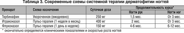
The duration of treatment with any drug depends on the clinical form of onychomycosis, the extent of the lesion, the degree of subungual hyperkeratosis, the affected nail and the patient's age. To calculate the duration, we are currently using our proposed special KYOTOS index. Combination therapy can be prescribed in cases where only systemic therapy is insufficient or it has a long duration. Our experience with terbinafine combination therapy includes its use in short courses and intermittent regimens, in combination with antifungal nail varnishes.
In the treatment of dermatophytosis of the feet and hands, both local and systemic antifungal agents are used. External therapy is most effective for erased and interdigital forms of mycosis of the feet ... Modern topical antimycotics include creams, aerosols, ointments. If these funds are not available, local antiseptics are used. The duration of treatment is from two weeks with modern drugs to four - when using traditional drugs. In case of chronic squamous-hyperkeratotic form of mycosis of the feet, involvement of hands or smooth skin, and nail damage, local therapy is often doomed to failure. In these cases, systemic drugs are prescribed - terbinafine - 250 mg per day for at least two weeks, itraconazole - 200 mg twice a day for one week. With nail damage, the duration of therapy is extended. Systemic therapy is also indicated for acute inflammatory phenomena, vesicular-bullous forms of infection. Outwardly, in these cases, lotions, antiseptic solutions, aerosols, as well as combined agents that combine corticosteroid hormones and antimycotics are used. Desensitizing therapy is indicated.
External therapy for lesions of smooth skin is indicated for isolated lesions of smooth skin. With the defeat of vellus hair, deep and infiltrative-suppurative dermatophytosis, tinea incognito, systemic therapy is indicated. We also recommend it for localization of foci on the face, and for widespread rubrophytosis (although, as a rule, nails are also affected).
Topical antifungals are used in the form of creams or ointments; use of aerosol is possible. The same drugs are used as for the treatment of mycosis of the feet. The duration of external therapy is 2-4 weeks. or until the disappearance of clinical manifestations and another 1 week. after. The drugs should be applied to the lesion and another 2-3 cm outward from its edges.
With simultaneous damage to the scalp or nails, systemic therapy is carried out according to the appropriate schemes. In other cases, with systemic therapy, terbinafine is prescribed at 250 mg / day for 2-4 weeks. (depending on the pathogen), or itraconazole with 1 cycle of pulse therapy (200 mg twice a day for 1 week). Similar schemes are used for inguinal dermatophytosis.
Prospects for combating dermatophytosis in Russia Currently, there is an almost continuous rise in the incidence of dermatophytosis. Most of the cases of the disease today account for mycoses of the feet and onychomycosis. At the same time, the official statistical picture of the incidence may differ from the real one, since a significant part of patients avoid going to medical centers.
Our studies of the etiology, epidemiology and clinical features of dermatophytosis showed that chronic infection caused by T. rubrum (so-called rubrophytosis) prevails in Russia ... Thus, most of the cases of dermatophytosis, at least in adults, is an anthroponous infection, and the only source of it is dermatophytosis patients themselves. At the same time, rubrophytosis, as shown by modern studies, including ours, is a disease with a long-term course, low severity of symptoms, and frequent intrafamilial transmission.
This raises the question of the feasibility of a complete victory over rubrophytic. The main goal of therapeutic and prophylactic measures should be the identification and treatment of patients with rubrophytosis. According to the set goal, we formulate the following tasks:
- Active search for patients with rubrophytosis ... This task can be carried out both within the framework of prophylactic medical examination programs and with the help of mass treatment and prevention campaigns of the “hot line” type. However, such methods are costly and cannot be implemented at the federal level. A more perfect approach to solving this problem can be effective sanitary and educational work focused on a constant flow of patients to specialized treatment centers. The introduction of programs for self-diagnosis of onychomycosis and mycosis of the feet, which increase the motivation for treatment, is promising.
- Improving therapies
... It is necessary to reach an acceptable low rate of relapses after treatment of onychomycosis, to improve and simplify treatment regimens, making them available not only to dermatologists, but also to general practitioners. From our point of view, the KYOTOS index, already introduced into clinical practice, is suitable for solving the last problem, which makes it possible to choose an adequate treatment regimen for onychomycosis and, at the same time, does not require significant clinical experience of the attending physician. To successfully combat rubrophytosis by general practitioners, it is also necessary to simplify and unify approaches to their laboratory diagnostics, sufficient to confirm the diagnosis. For this, direct PCR probes can be used to detect T. rubrum in clinical material, and work in this direction is already underway.
It is also necessary to find a compromise between the cost and effectiveness of treatment, for which a combination therapy with keratolytics can be used to avoid prolonged courses of systemic therapy.
- Development of fundamentally new means of prevention ... The immediate task is sanitary and educational work aimed at early prevention and prevention of rubrophytosis before the development of onychomycosis, the treatment of which is associated with great difficulties and costs. Further research is needed to clarify certain aspects of the proposed strategy for combating dermatophytosis, work in the existing directions. And all the more, it is necessary to unite the efforts of specialists and scientists, practicing doctors and healthcare organizers of different profiles.
1. New in the taxonomy and nomenclature of mushrooms. Under. ed. Yu. T. Dyakova and Yu. V. Sergeeva. M .: 2003.164-192.
2. Sergeev A. Yu., Sergeev Yu. V. Fungal infections. A guide for doctors. Moscow: BINOM-Press, 2003.440 p.
3. Sergeev A. Yu., Ivanov O. L., Sergeev Yu. V., Vakhlakov A. N., Sedova T. N., Dudnik V. S. Research of modern epidemiology of onychomycosis. Bulletin of Dermatology and Venereology. - 2002; 3: 31-35
4. Sergeev A. Yu. Modern ideas about the pathogenesis of onychomycosis. Immunopathology, allergology, infectology. 2000; 1: 101-110.
5. Potekaev NN Microsporia. Russian medical journal. 2000; 8 (4): 189-196.
6. Sergeev Yu. V., Sergeev A. Yu. Onychomycosis. Fungal nail infections. M .: Geotar medicine. 1998, 126 p.
7. Sergeev A. Yu. Fungal diseases of nails. M .: National Academy of Mycology - Medicine for all. 2001.- 164 S.
8. Sergeev Yu. V., Sergeev A. Yu., Mokina EV Buchinsky OI Hotline: The first mass campaign to identify and treat patients with onychomycosis. In the book: advances in clinical immunology and allergology. (under the editorship of A. V. Karaulov). M .: 2002.- S. 355-363.
9. Guidelines for laboratory diagnosis of onychomycosis. Ed. A. Yu. Sergeeva. M .: Geotar medicine. 2000, 154 p.
10. Sergeev Yu.V., Potekaev NS, Leshchenko VM, Larionova VN .. Lamisil: improving the therapy of onychomycosis caused by dermatophytes // Bulletin of dermatology and venereology. 1995; 5: 54-56.
11. Potekaev NS Kurdina MI, Potekaev NN Lamisil with microsporia. Vestn. dermatol. 1997; 5: 69.
12. Sergeev Yu. V., Sergeev A. Yu., Leshchenko VM Modern program of fight against dermatomycosis in Russia. In the book: Advances in medical mycology. (under the editorship of Yu.V. Sergeev) M .: 2002.Vol. 2.P. 160-162.
Dermatophytosis is a common dermatological disease. The pathology has an infectious etiology. The causative agents of the disease are fungi belonging to a large subclass of dermatophytes.
Dermatophytosis is of several types. There are keratomycosis, epidermophytosis inguinal, rubrophytosis, athlete's foot, favus, tartar mycosis, histoplasmosis, candidiasis, erythrasma, etc.
The disease can only be eliminated by medication. It is pointless to make any surgical interventions. To eliminate the inflammatory process and suppress the vital activity of fungi, antifungal drugs are used.
What is this article about?
Pathogens and causes of the disease
Dermatophytosis of smooth skin is an infectious disease. Pathology cannot develop due to unbalanced diet, bad habits, bad heredity - these are all myths.
In fact, bad habits can only be attributed to predisposing factors, but not to the main cause of the development of the disease. So, why does dermatophytosis develop and who is its causative agent?
The main cause of the disease is invasion by pathogenic microorganisms. Often, fungal microorganisms are transmitted by the household route - through a towel, common kitchen items, hygiene products.
The causative agents of dermatomycosis can be divided into three main groups, including:
- Microsporum.
- Epidermorhyton.
- Trichophyton.
Doctors note that 26-30 degrees Celsius is considered a favorable temperature for the reproduction of these microorganisms, therefore, dermatomycosis develops much more often in people living in a tropical climate.
Immunity plays an important role. If a person has a malfunction of the immune system, then the fungal disease will be more difficult. Moreover, immunodeficiency often leads to chronicity of the infectious process.
Unbalanced diet, bad habits, hormonal disorders, diabetes mellitus, excessive sweating and overweight predispose to dermatophytosis.
Types of dermatophytosis
Inguinal
Inguinal dermatophytosis is uncommon. According to statistics, men are most susceptible to this disease. A fungal infection is transmitted during sexual intercourse. Moreover, an interesting fact is that even barrier contraception may not protect against illness.
The fact is that dermatophytes most often "live" on the pubis. Condoms do not protect a person from infection in the skin folds, so condoms are not a 100% guarantee that a person is completely protected.
Inguinal dermatophytosis manifests itself in the following symptoms:
- The appearance of a rash in the groin area. Sometimes the perineum and anus are affected. Usually, the spread of the rash occurs in the absence of timely treatment.
- Itching in the genital area.
- Local burning.
- Tenderness to palpation.
Primary foci of mycosis outwardly resemble pink spots with clear boundaries. Over time, the spots begin to merge and form large foci. Scales, crusts, various bubbles and even suppuration can form.
In the absence of timely treatment, the fungal infection begins to spread throughout the body.
Mycosis of feet and hands
Dermatophytosis of the feet, interdigital space and hands is very common. Usually, this disease develops due to non-compliance with hygiene rules, or contact with the personal belongings of the infected.
Almost all dermatophytes, including Candida, microorganisms from the genus Malassezia can be the causative agents of mycosis of the feet / hands. Fungus of the feet is rarely accompanied by damage to the deep layers of the dermis - they, as a rule, remain "intact".
The characteristic signs of the disease are:
- Thickening and increasing dryness of the skin. On the feet, the skin can thicken, which is called "rough calluses." Cracks often form, the likelihood of secondary infections increasing.
- Lamellar or mucous peeling of the skin at the site of the lesion.
- Leukonychia. Doesn't always appear. This term means a phenomenon in which white stripes or spots form on the nail plates.
- Itching and burning.
- Discoloration of the dermis at the site of the lesion.
- Hyperemia (local increase in body temperature).
- The appearance of dry papules. The occurrence of cyanotic plaques is possible.
With the defeat of the feet / hands, in 95% of cases, the fungal infection spreads to the nail plate.
Dermatomycosis of the scalp
 Dermatophytosis of the scalp occurs quite often in children. The causative agents of the disease get on the skin upon contact with the things of an infected person. But it is possible to "catch" the disease by contact with infected animals or from the soil.
The immune factor plays an important role. Dermatophytosis of the head most often develops with a reduced level of immunity. This explains the widespread prevalence of an infectious disease among children.
Symptoms of mycosis:
- The appearance of foci of alopecia (baldness) on the head.
- Increased hair fragility.
- Dandruff.
- The appearance of blackheads in areas of baldness.
- Increased hair oiliness.
- Peeling of the skin.
In the acute course of the infectious process, purulent foci appear, mucous exudate is separated, and crusts form. The lesions can reach 4-5 centimeters in diameter.
Nail fungus
The nail is affected by dermatophytes very often, especially if the infection has already spread to the palms, interdigital space, feet, hands. The defeat of the nail plates is the easiest to treat, and, moreover, has the most pronounced symptoms.
The main manifestation of a fungus is a discoloration of the nail. It usually turns yellow or brown. In some cases, the nail plate turns black, green, or white.
In addition to discoloration of nails, an infected person has:
- The appearance of abscesses near the nail bed.
- Deformation of the nail plates. They begin to exfoliate, crumble easily, change their shape. Often the nail plates grow in and must be removed surgically.
- Itching and burning.
- Emergence
- Thinning of the nail plates.
In the absence of proper treatment, the nails ultimately atrophy and their complete destruction occurs.
How to treat dermatophytosis?
Traditional methods
With dermatophytosis of the nail, treatment can be done at home. Alternative methods are no less effective for fungal infections of the inguinal folds, scalp, feet, hands, interdigital space.
The list of effective remedies for fungus includes:
- Compresses with aloe juice. Apply to the affected area 2-3 times a day.
- A decoction of calendula and mother-and-stepmother. Recommended to be taken orally to enhance immunity. The optimal daily dose is 150 ml.
- Leuzea tincture. It is a powerful immunostimulant, has antiseptic and antifungal effects. It is recommended to take 10-15 drops 3 times a day.
- Soda baths. It is recommended to take in case of nail fungus, feet, interdigital space, palms. It is enough to add 3-4 tablespoons of baking soda to warm water, then take a bath for 20-30 minutes. Repeat the procedure 4-5 times a day.
- Tea tree oil baths. Add about 100-150 ml of oil to the water. Take a warm bath for at least 20 minutes. Repeat the procedure 3 times a day.
- Lotions with hydrogen peroxide. This remedy was recommended for use by Dr. Neumyvakin. According to the professor, hydrogen peroxide is able to destroy almost any dermatophyte. The physician recommends moistening a cotton swab in peroxide, and then applying it to the affected lesion for 10-15 minutes. The procedure should be repeated 2-3 times daily.
Before using folk remedies, a doctor's consultation is required, since the above drugs, under certain conditions, can be harmful.
Drug therapy
Dermatophytosis is usually treated conservatively. Without fail, patients are prescribed antifungal medications with a pronounced fungicidal and fungistatic effect.
In the treatment of children and adults, capsules, and ointments for external use, and antifungal varnishes, and sprays can be used. Specific drugs are selected based on the type of mycosis and the type of pathogen. To find the right products, it is imperative to pass a scraping for fungus.
Consider each form of drug release separately:
- Capsules. Mostly used for lesions of smooth skin, scalp, groin. Much less often prescribed to persons suffering from fungus of feet, nails, palms. The most effective capsules are Fucis, Itracon, Nystatin, Diflucan, Irunin, Itraconazole, Clotrimazole.
- Ointments, gels, creams. Usually prescribed for interdigital space, nails. Judging by the reviews, the most inexpensive and effective drugs in this segment are Clotrimazole, Exoderil, Nitrofungin, Lamisil, Mikoseptin.
- Sprays. They are used in the treatment of dermatomycosis of the legs and nail plates. The most effective aerosols are Lamisil, Thermikon, Lamikon.
- Healing varnishes. Used to treat nail fungus. The most effective are Batrafen, Oflomil, Lotseril. During the period of using varnishes, it is prohibited to do manicure / pedicure. Antifungal drugs in this segment are allowed to be used for prophylactic purposes.
Along with antifungal medicines, antiseptics, immunomodulators and multivitamin complexes can be used. Assigned optionally.
Excess heat and moisture creates a favorable environment for mushroom growth.
Dermatophytes are spread by contact with infected animals or people, as well as contaminated household items.
Dermatophytosis of smooth skin is a common superficial fungal infection of smooth skin characterized by well-defined annular lesions with central resolution, erythema, and peripheral peeling.
Dermatophytes: Trichophyton, Microspornm and Epidermophyton spp.
Diagnostics of the dermatophytosis of smooth skin
Diagnosis established on the basis of anamnesis, examination and microscopy.
Morphology: well-defined annular lesion with central resolution, erythema and desquamation along the periphery. Concentric lesions are highly specific (80%) for dermatophyte infections.
Other characteristic signs: itching in the affected area.
The lesions can be located anywhere on the body, including the face and underarms.
Unrecognized or tinea incognito is a dermatophytic infection that has not been previously recognized by the doctor / patient, and topical steroids have been used to treat the lesion. When steroids are used, the dermatophyte continues to grow, creating cosmetic problems. In some cases, the infection causes hyperpigmentation.
Dermatophytosis of smooth skin can invade large areas of the body.
KOH microscopy is useful for confirming clinical findings or when the diagnosis is unclear. For this purpose, a scraping is taken from the peripheral and erythematous area of \u200b\u200bthe lesion, using the edge of a glass slide or a scalpel. To obtain a sufficient amount of the stratum corneum without causing bleeding, the procedure must be performed with pressure. If the material is not taken correctly, as well as in cases where the patient uses local antifungal drugs or microscopy is performed by an inexperienced specialist, a false negative result can be obtained.
For faster dissolution of epithelial cells without heating, KOH with dimethyl sulfoxide (DMSO) is used. Fungal dye can be used.
Skin scraping with culture analysis is the gold standard, but is more expensive and can take up to two weeks to grow.
If the KOH test and culture are negative, and the clinical picture still indicates a fungal infection, a biopsy should be performed by sending the obtained material in formalin to a laboratory for Schiff staining.
Differential diagnosis of skin dermatophytosis
Granuloma annularis is an inflammatory benign dermatosis of unknown origin, which is characterized by both dermal and annular papules.
Psoriasis is characterized by plaques with scales on the extensible surfaces of the trunk. Sometimes the plaques are annular. Inverse psoriasis in intertriginous zones can also mimic smooth skin dermatophytosis.
With erythema annular centrifugal, flaky red rings appear with a patch of normal skin in the center, with the desquamation following the erythema as the annulus expands, while with dermatophytosis, the desquamation goes ahead of the erythema.
When infected with a cutaneous migratory larva, serpiginous passages are observed, laid by the larva of the crooked head, which can have a ring-shaped pattern and be mistaken for dermatophytosis of smooth skin.
Numular eczema is characterized by rounded, coin-shaped, red, scaly plaques without permission in the center.
Erythrasma is localized in the axillary and groin areas, does not have an annular configuration and does not have a resolution in the center. Coral-red underneath Wood's lamp.

Treatment of dermatophytosis of the skin
In case of dermatophytosis in limited areas of smooth skin, local antifungal drugs are used.
Although almost all topical antifungal agents are effective in the treatment of dermatophytosis of the feet and smooth skin, clinical data indicate a better efficacy of allylamines (terbinafine) compared to expensive azoles.
Research shows that 1% terbinafine cream or solution (once daily for seven days) is highly effective for smooth skin dermatophytosis and groin dermatophytosis. With 1% cream (known commercially as Lamisil), mycological efficacy was 84.2%, compared with 23.3% with placebo.
The average number of courses required for treatment was 1.6.
If dermatophytosis of smooth skin occupies large areas of the body, systemic antifungal drugs are considered first-line therapy. However, if the size of the affected area is limited, it will not be a mistake to try local therapy. A patient with unrecognized dermatophytosis required systemic therapy to resolve the infection. Unfortunately, post-inflammatory hyperpigmentation has not completely resolved.
A randomized control study showed that in the treatment of smooth skin dermatophytosis and inguinal dermatophytosis, itraconazole 200 mg orally given daily for one week was as effective, safe and well tolerated as itraconazole 100 mg for two weeks.
In one study, patients with a laboratory diagnosis of smooth skin dermatophytosis and inguinal dermatophytosis were randomly divided into two groups, which received either 250 mg terbinafia once a day or 500 mg griseofulvin once a day for two weeks. The efficacy for terbinafia was higher at week six.
So, if a systemic drug is needed, clinical evidence supports the use of
- Terbinafia at a dose of 250 mg daily for two weeks,
- Itracoiazole 200 mg daily for one week,
- Itracoiazole 100 mg daily for two weeks.
The patient is advised to keep the skin dry and clean. Infected pets must be treated.
In case of difficult to treat and widespread disease, a second visit to the doctor is scheduled in 4-6 weeks. If bacterial superinfection is likely, the control examination should be carried out earlier.
Clinical example of smooth skin dermatophytosis... A six-year-old girl was brought to the doctor for a round, itchy lesion on her body. The rash was first discovered two weeks ago. The domestic cat has had several areas of hair loss. Notice the concentric circles with scaling, erythema, and center resolution. Under Wood's lamp, the focus glowed green; analysis with KOH revealed branching and septic hyphae. The child was prescribed a local antifungal cream twice a day, and after 3-4 weeks the dermatophytosis resolved.
Dermatophytosis is an extremely common skin disease, the diagnosis of which causes some difficulties associated with the similarity of the disease with other skin pathologies. Treatment requires radical methods and a fairly long period of time.
The causative agents of dermatophytosis are molds of the Arthodermataceae family. In total, they have 43 varieties, 30 of which are capable of causing skin diseases.
Increasingly, scientists are calling fungal infections the plague of the twenty-first century. And this comparison has serious grounds:
- the spread of infection is extremely rapid;
- drug methods of treatment today still leave much to be desired;
- all forms and types of fungal diseases have not yet been identified.
The main types of dermatophytosis
Dermatophytosis has many forms and varieties, which causes certain difficulties in its diagnosis. It is possible to recognize the source of tissue damage only in laboratory conditions; a superficial examination of the skin, as a rule, does not give any results.
The types of the disease are distinguished depending on the following factors:
- The type of tissue affected.
- Localization of the lesion.
Definition by the type of tissue affected
- epidermophytosis: the stratum corneum of the epidermis is affected;
- trichophytosis: the stratum corneum of the epidermis and scalp are affected;
- onychomycosis: the nail plates are affected.

information to read
Determination by localization of the lesion
Fungal infections can affect the skin of the following areas of the body:
- feet and hands;
- groin area;
- smooth skin and large folds;
- torso.
Accordingly, the disease is classified according to the location of the lesion. For example, smooth skin dermatophytosis or inguinal dermatophytosis.
Note. There is another type of disease: undefined dermatophytosis. As the name suggests, it does not have a specific target area.

Dermatophytosis of the feet
Dermatophytosis of the feet is the most common disease among all types of dermatophytosis. According to statistics, approximately 70% of all people on the planet at a certain period of their lives were infected with the causative agent of a fungal infection.
Foot fungus has the following features:
- rarely occurs in childhood (pre-adolescence);
- more often men suffer from the disease than women.

Types of dermatophytosis of the feet:
- Interdigital dermatophytosis... The foci of development are observed in the interdigital spaces, in neglected forms they pass to the sole. There is a dry type of disease, in which peeling predominates, and a wet type, characterized by constant maceration, that is, wetting of the affected surfaces.
- Moccasin dermatophytosis... Manifested by severe peeling of the plantar and lateral parts of the feet. The scales are white or greenish.
- Inflammatory form of dermatophytosis characterized by the appearance of blisters and vesicles on the affected skin surfaces.
Important! It is worth noting that with any form of dermatophytosis of the feet, there is a strong, almost unbearable itching, which only intensifies when scratching, and the affected areas quickly turn red.

Inguinal dermatophytosis
Basically, this type of fungal disease manifests itself in men, less often in women and children. The main signs are redness, peeling, and severe itching. In advanced cases, blisters appear. As a rule, skin irritation is localized in the groin area. However, lesions often extend to the abdomen and peri-groin areas of the thighs.
The clinical picture is red scaly plaques of various sizes. Sometimes they look like lichen, growing one inside the other.
If untreated, groin dermatophytosis can last from several months to several years.

Important! Often, inguinal dermatophytosis accompanies a fungal infection of the feet, since in most cases the causative agent of the infection is carried by hands from the feet to the groin area. This factor is decisive when a disease occurs, when there is no basic hygiene.
This type of disease is distinguished by a specific clinical picture, therefore, in most cases, a dermatologist can detect it even with a superficial examination.... But the disease sometimes looks very much like the manifestations of erythrasma. In this case, the diagnosis is carried out using a Wood lamp.

Dermatophytosis of the hands
Dermatophytosis on the hands is a fairly common type of disease. Fungal rash on the hands is localized, as a rule, on the dominant hand, and is chronic. The type of rash is papules and vesicles. As with other types of dermatophytosis, severe itching is observed, especially when water gets on the affected areas. When scratching, bloody discharge may occur, turning into purulent wounds, if the correct treatment is not applied.
Localization sites are the inner parts of the palms and the lateral surfaces of the fingers. In addition to the appearance of papules, there is reddening of the skin in clearly limited areas, as well as constant peeling.
Note. Dermatophytosis of the hands brings patients special inconvenience, since these areas of the skin are constantly in contact with water, which increases discomfort and causes exacerbation of the disease.

Dermatophytosis of smooth skin
Until recently, it was not recognized and classified by scientists as a separate type of fungal disease. Its external signs resemble other types of dermatophytosis, however, they do not have specific localization sites, and, as a rule, arise on areas of the skin on which hair does not grow. These are the neck, abdomen, inner surfaces of the limbs, back, some areas of the face, for example, the temples or cheeks.
This type of disease often affects large folds of the skin. This is the groin area, the back of the knees and elbows.

Diagnostics of the dermatophytosis
The question “What is dermatophytosis and how to treat it?” Can be answered only after laboratory examination of scrapings from the affected skin areas. Microscopic examination of materials such as skin, nails, hair, you can see fungal masses, which are the causative agents of the disease.
Diagnosis of all types of dermatophytosis requires the same procedures. It:
- collection of anamnesis;
- external examination of the patient;
- sometimes - inspection with a Wood lamp.
Important! Some types of dermatophytosis are easy to confuse with other types of skin diseases, therefore, treatment must be started with a qualified diagnosis, otherwise it is possible to achieve only aggravation of the patient's condition.

The main method for diagnosing dermatophytosis is laboratory examination of skin samples for the presence of fungal microorganisms
Dermatophytosis treatment
It should be noted that almost all types of dermatophytosis are treated in the same way. The specialist prescribes a course of antifungal therapy. In this case, only the degree of neglect of the disease matters. Depending on this, a course of antifungal (antimycotic) drugs and / or antibiotics may be prescribed.
Local antibacterial and antifungal agents are mandatory. If the affected skin gets infected, antiseptic agents should also be used.
The most effective topical remedies for dermatophytosis include a 1% solution or Terbinafine cream. The recommended course of treatment is 2-3 times a day for a week.

Treatment of dermatophytosis should be comprehensive, one of its components is the use of local remedies
If it is necessary to use systemic drugs, experts recommend the following funds:
- Terbinafine;
- Itraconazole.
It is imperative to keep the skin clean and dry. If there are infected animals in the house, they need to be cured. If the treatment does not bring the desired results, it is recommended to re-check with the doctor after 4-6 weeks.
Prevention of dermatophytosis
Undoubtedly, dermatophytosis is an infectious disease characterized by a fairly high degree of infection.

Compliance with hygiene standards is the most important condition for preventing dermatophytosis
However, its development can be prevented by adhering to basic hygiene standards:
- using only clean underwear and bed linen, including towels;
- taking a shower or bath every day;
- frequent hand washing;
- daily change of underwear and socks;
- after water procedures, it is recommended to wipe your feet dry.
Important! Fungal microflora loves a humid and warm environment. Therefore, it is necessary to monitor the condition of shoes, especially closed ones, in a warm period.
It is worth remembering that the main causes of dermatophytosis are non-compliance with hygiene standards, therefore, you should be especially careful about such moments as visiting public places (baths, swimming pools, etc.). After all, almost everyone can "pick up" the fungus.
It should not be assumed that dermatophytosis of the trunk is some kind of specific disease, since this is a whole group of diseases that are caused by various fungi, can manifest themselves with different symptoms, are treated differently and have different medical and social significance.
The unifying factor is that all dermatophytosis of the trunk have a characteristic localization (they do not include fungal infections of the scalp, groin folds, palms, feet and nails), and are also caused by fungi, and not by other microorganisms (bacteria, viruses, protozoa) ...
Why does a fungal infection occur?
- Trichophyton rubrum;
- Trichophyton mentagrophytes;
- Microsporum canis;
- Microsporum gypseum.
Less commonly, the disease is caused by other fungi. At the same time, some of the microorganisms can only lead to damage to the scalp, for example, and does not cause epidermophytosis.
But in addition to the actual cause of the disease, there are also a number of predisposing factors. We all know that fungal diseases are observed, although often, but not in everyone, although almost all people are in contact with the pathogen.
Predisposing factors that can contribute to the development of fungal skin lesions are:
- Decreased immunity due to malnutrition, chronic stress, congenital immunodeficiency or in the presence of HIV;
- Inadequate skin care, excessive sweating and a shift in pH towards the acidic side can cause the fungus to more easily penetrate the skin and spread quickly;
- The presence of skin lesions through which the pathogen penetrates;
- Failure to comply with the rules of personal hygiene when visiting public places (baths, saunas, gyms, swimming pools);
- Contact with infected outdoor animals or pets that have been infected with a fungal infection.
Signs of mycosis on the skin of the trunk
We figured out what dermatophytosis of the trunk is, and now it is necessary to understand exactly what symptoms this disease can manifest. Certain clinical manifestations may vary depending on the causative agent of the disease. Different fungi lead to the appearance of different symptoms, but the differences are often so insignificant that even a doctor cannot distinguish them during a detailed examination, therefore, for the final confirmation of the diagnosis, appropriate examinations are necessary.


And yet, dermatophytosis of the body is manifested by a number of general symptoms, among which should be called:
- The appearance of a spot of redness on the skin;
- The presence of single or multiple spots;
- In the center of the spot, less pronounced inflammation is always observed (manifested by reddish and flaky skin with dermatophytosis of the trunk), and more pronounced along the edges;
- The edge of the spot is elevated, it can be covered with papules or pustules;
- Peeling of the skin is observed, mainly along the edges;
- The lesion may gradually increase;
- In some cases, itching is observed, in others - the spot may not bother in any way;
- There is no pain at the site of the lesion, so the stain may remain unnoticed for a long time.
Diagnostics
Dermatophytosis of the trunk should be distinguished from many other diseases that can also manifest as foci of redness and flaking of the skin, among them:
- bacterial skin inflammation;
- annular erythema;
- in some cases with.
Only the clinical picture does not always make it possible to carry out a differential diagnosis, despite the fact that the dermatophytosis of the trunk of the spots can be localized anywhere, while in psoriasis they have a clear localization, and in neurodermatitis, for example, there is a pronounced subjective symptomatology that is not present in dermatophytosis.
To make an accurate diagnosis, it is not enough to assume simply the presence of dermatophytosis of the trunk, since the type of pathogen is also of great importance for the correct selection of therapy. Therefore, scraping is always performed from the affected area. Microscopic examination of horny scales allows you to detect hyphae of a particular fungus and make an accurate diagnosis.
Treatment of dermatophytosis of the trunk
Treatment of fungal skin lesions involves the use of local and systemic drugs. Local remedies - ointments and creams with antifungal components. The most widely used drugs are the azole group (the most famous of them is ketoconazole), but local therapy is quite effective only in the case of an acute course of skin mycosis.
In chronic dermatomycosis, the use of systemic drugs is always necessary. Often, drugs with a wide spectrum of action are used, but in some cases they turn out to be ineffective, but drugs that are narrower in their spectrum provide a better clinical effect. Therefore, the decision on the appointment of a particular therapy should be made after a full examination and an accurate diagnosis, and not on the basis of only a clinical picture without any diagnosis.
The use of only antifungal drugs (both local and systemic) may not bring results in the case of pronounced suppression of immunity, in the case of a large amount of sweets in the diet (shift the pH of the skin to the acidic side) and the action of some other factors.
In this regard, treatment should always be prescribed by a doctor, be complex and based on the nature of skin lesions (one or another type of pathogen of dermatomycosis), self-medication may be not only ineffective, but also dangerous.
Traditional methods of treatment
It turns out to be quite difficult to deal with dermatophytosis of the trunk using folk methods. If even specialized ointments containing antifungal components do not help with many forms of this disease, then what can we say about folk remedies that have a predominantly anti-inflammatory effect.
For the most part, folk recipes are aimed not at the cause (fungal infection), but at the symptoms of the disease (redness and itching). The anti-inflammatory effect of chamomile, calendula, fir and oak bark allows the use of decoctions and infusions from these components for the treatment of dermatophytosis.
Mostly baths and compresses are used, but the isolated use of such approaches cannot be recommended, since, despite some reduction in symptoms, the cause cannot be eliminated. Traditional methods are used exclusively as an adjunct to the main therapy in the absence of contraindications (first of all, we are talking about allergic reactions).
Prevention
Measures to prevent dermatophytosis of smooth skin are reduced to measures to prevent contact with the fungus and increase immunity. These should include:
- Strict adherence to hygiene rules when visiting public institutions (use of personal shoes and towels);
- Proper skin care, regular linen change, combating excessive sweating;
- Compliance with the regime of work and rest, proper nutrition, rejection of bad habits.
The key to effective secondary prevention of dermatophytosis of the trunk is the timely identification of lesions, a full examination for the correct diagnosis and complex treatment.
Photo


